The human spine is a complex and vital structure in our body, playing several crucial roles, including supporting the body’s weight, providing flexibility for movement, and protecting the spinal cord. Understanding the basics of spinal anatomy can help you grasp the importance of spinal health and the implications of spinal disorders. This blog aims to demystify spinal anatomy, by breaking it down into its essential components and their functions.
The Basics of The Spine
The spine, also known as the vertebral column or backbone, is a column of 33 individual bones called vertebrae, which are stacked one on top of the other. These vertebrae are categorized into five regions: cervical, thoracic, lumbar, sacral, and coccygeal. Each region has a different number of vertebrae and serves distinct functions.
1. Cervical Spine: Your Neck’s Foundation
- Vertebrae: C1-C7
- Key Roles: The cervical spine holds up your head, which might weigh between 10 to 12 pounds (about the weight of a bowling ball!). It also allows for a wide range of head movements—up and down, side to side, and rotation. This flexibility makes it prone to injury, so it’s crucial to maintain good posture and strengthen neck muscles.
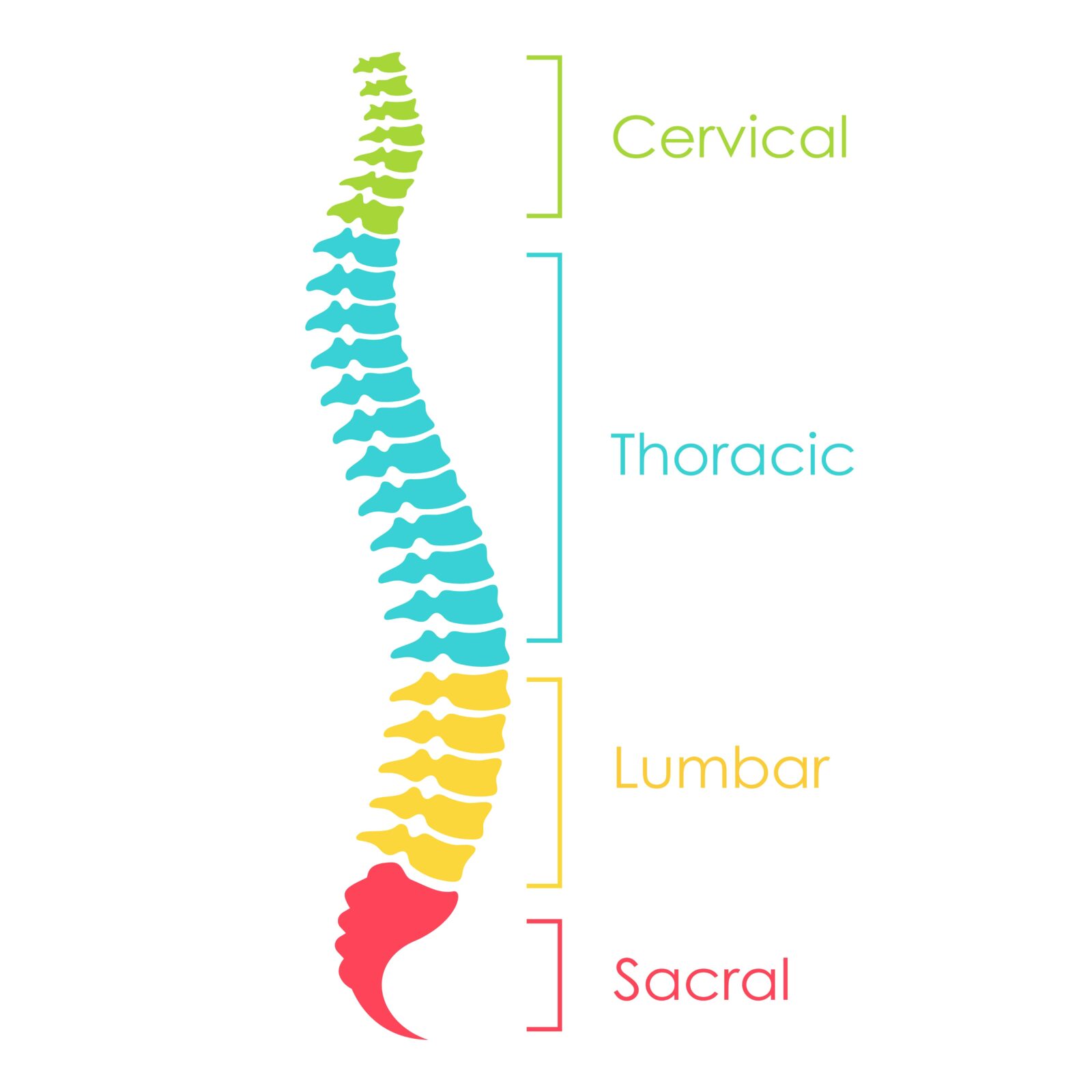
2. Thoracic Spine: The Upper Back Anchor
- Vertebrae: T1-T12
- Key Roles: This region is your upper back, where your ribs attach to the spine, forming the back part of your rib cage. The thoracic spine plays a pivotal role in providing stability and protection for your vital organs. Its range of motion is more limited compared to the cervical and lumbar areas, mainly because of its role in safeguarding the heart and lungs.
3. Lumbar Spine: The Lower Back Workhorse
- Vertebrae: L1-L5
- Key Roles: The lumbar spine bears the brunt of your body’s weight, especially when lifting, bending, and twisting. It’s designed to be both strong and flexible, but this combination also makes it susceptible to injury. Keeping your core muscles strong can help support this region, reducing the risk of lower back pain.
4. Sacral Region: The Stability Core
- Vertebrae: 5 fused vertebrae forming the sacrum
- Key Roles: The sacrum sits between your hip bones, connecting the spine to the pelvis. This region’s main job is to provide stability for the body by distributing weight evenly across the pelvis and legs. The sacrum also forms the back wall of the pelvic cavity, playing a key role in pelvic organ support.
5. Coccygeal Region: The Tail End
- Vertebrae: 3-5 fused vertebrae forming the coccyx (tailbone)
- Key Roles: The coccyx, or tailbone, is the final segment of the vertebral column. It serves as an attachment site for tendons, ligaments, and muscles of the pelvic floor. While it may seem small and insignificant, injuries to the coccyx can be quite painful and take a long time to heal.
Each of these regions has a specific structure and function, contributing to the spine’s overall role in your body. From providing mobility to ensuring stability and protecting the spinal cord, the diverse functions of these spinal regions highlight the complexity and importance of spinal health.
Anatomy of a Vertebra
Each vertebra in your spine is a marvel of natural engineering, designed to provide support, flexibility, and protection to your spinal column. Despite the slight variations to fit their specific functions in different regions of the spine, all vertebrae share a common structural anatomy. Let’s delve into the anatomy of a typical vertebra to understand how these critical components of your spine work together.
Vertebral Body
The vertebral body is the thick, disc-shaped anterior (front) portion of a vertebra. It’s the main weight-bearing structure, designed to support the body’s weight and withstand compressive loads. The strength of the vertebral bodies is crucial for maintaining posture and providing a stable platform for the rest of the body. The size of these bodies increases from the cervical to the lumbar region to accommodate the increasing load as you move down the spine.
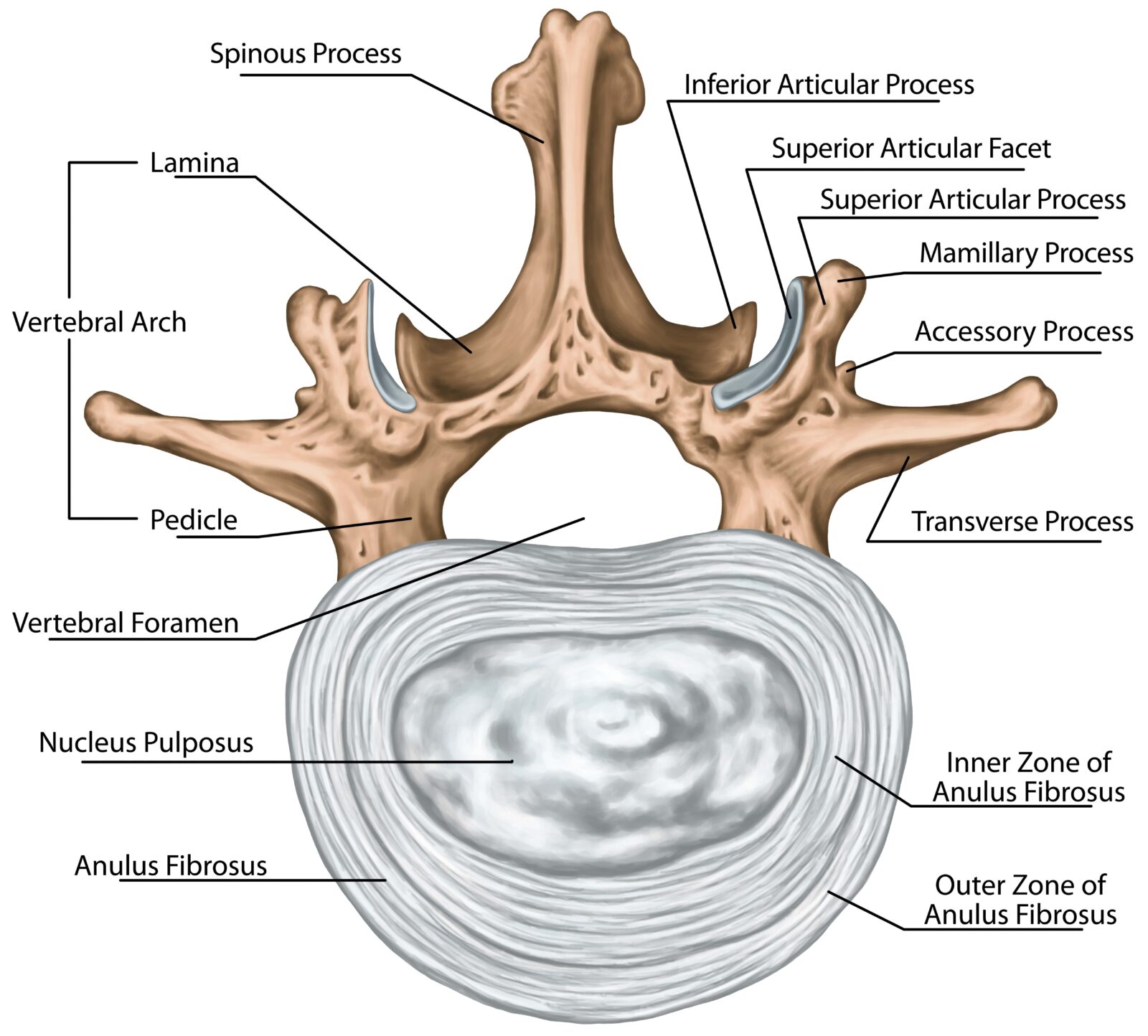
Vertebral Arch
The vertebral arch forms the posterior (back) part of the vertebra. It consists of two pedicles and two laminae, creating a ring-like structure that encloses the vertebral foramen. The vertebral arch protects the spinal cord by forming the vertebral foramen, the canal through which the spinal cord passes. It also serves as a point of attachment for muscles and ligaments.
Spinous Process
The spinous process is a bony projection that extends backward from where the two laminae meet. It’s what you can feel under the skin along the middle of your back. This process serves as an attachment point for muscles and ligaments. It plays a key role in movement and flexibility of the spine.
Transverse Processes
Located on either side of the vertebral arch, these are small bony projections where the laminae join the pedicles. Transverse processes serve as levers and attachment points for muscles and ligaments, aiding in the spine’s movement and stability.
Articular Processes
Each vertebra has four articular processes (two superior and two inferior) that protrude from the vertebral arch. These processes form joints with the articular processes of the vertebrae above and below. These joints, known as facet joints, guide and restrict the spine’s movement, preventing excessive rotation or flexion that could damage the spinal cord.
Intervertebral Foramina
Not a part of the vertebra itself but formed between two adjacent vertebrae, these are openings that allow for the exit and entry of spinal nerves. These foramina are critical for the nervous system, providing pathways for spinal nerves to connect the spinal cord with the rest of the body. Any obstruction or narrowing of these openings can lead to nerve pain or damage.
Intervertebral Discs
Between each vertebral body lies an intervertebral disc, which is not a part of the vertebra but plays a crucial role in the spine’s function. These discs act as shock absorbers, cushioning the vertebrae and allowing for flexibility. Each disc has a tough outer layer (annulus fibrosus) that surrounds a gel-like center (nucleus pulposus), providing the perfect balance between strength and elasticity.
The Support Crew: Ligaments, Tendons, and Muscles
Beyond the vertebrae and intervertebral discs, the spine’s integrity and functionality depend heavily on a network of ligaments, tendons, and muscles. This support system not only stabilizes the spine but also facilitates movement and protects against injury. Let’s explore the roles of these critical components.
Ligaments: The Stabilizers
Ligaments are tough, fibrous tissues that connect bones to other bones. In the spine, they play a pivotal role in maintaining the alignment of the vertebrae and limiting excessive movement that could lead to injury.
Tendons: The Connectors
Tendons are similar to ligaments but connect muscles to bones. In the spine, tendons attach the spinal muscles to the vertebrae, facilitating the transfer of force as muscles contract and relax. This action enables the spine to move in various directions while maintaining its stability and alignment.
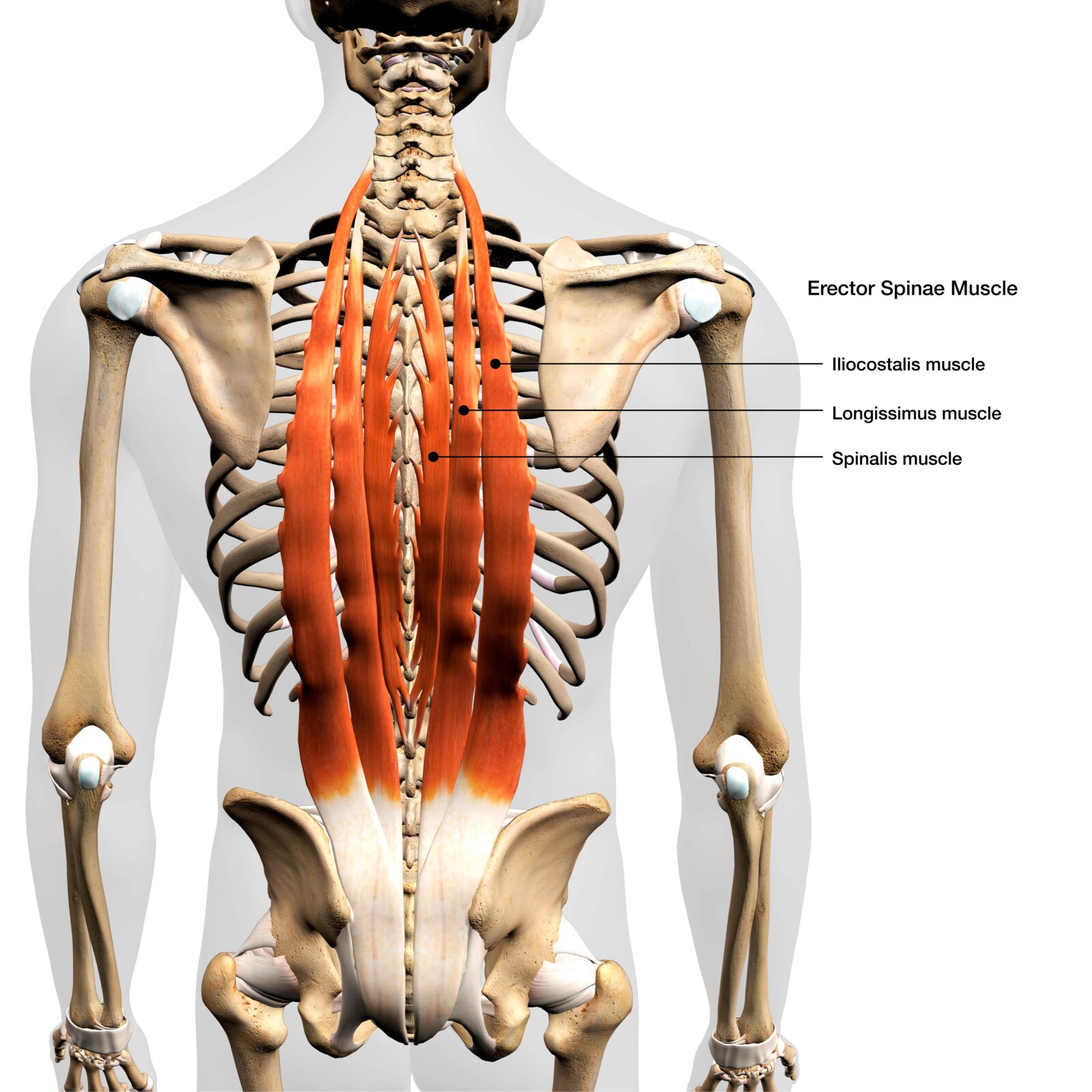
Muscles: The Movers
The muscles of the back are categorized into two main groups based on their location and function: the intrinsic (deep) muscles, which are primarily responsible for the movement and stability of the spine, and the extrinsic (superficial) muscles, which are involved in larger movements of the trunk and limbs.
In Conclusion: Your Spine, Your Health
Your spine is a central part of your well-being, playing a crucial role in your daily life. By understanding its basic structure and function, you’re better equipped to take care of it. From the intricate structure of each vertebra to the vital roles of intervertebral discs, ligaments, tendons, and muscles, the spine is a marvel of natural engineering that supports our body, enables movement, and protects vital neural pathways. By familiarizing ourselves with the components of the spinal column and their functions, we can better understand how to care for our spine through proper posture, regular exercise, and healthy lifestyle choices. Recognizing the signs of potential spinal issues and seeking timely medical advice can also help in maintaining spinal health. Ultimately, understanding spinal anatomy empowers us to take proactive steps towards preventing back pain and other spinal conditions, leading to a more active, healthy, and fulfilling life.